
解答は A〜E のいずれか1つ
1 / 2
標本・図の同定、神経支配、位置関係を、大学の実技・口頭試験に合わせて対策します。
頭蓋、下肢、上肢を写真で観察。英語の問題に答え、日本語・英語の解説で位置と働きを確認。
目印を押すと拡大する位置が切り替わる。部位名と説明は解答後に表示。
解剖模型の写真 / Anatomical model photograph
図版:Bruce Forciea (PhysioAnatomica) · CC BY-SA 4.0
Bruce Forciea (PhysioAnatomica)
図版の出典 · CC BY-SA 4.0
原画像の構造・色・背景は変更せず、表示用に縮小してWebPへ変換。 目印・解説・問題はMIRUNOTEが追加。改変図版の利用条件は上記ライセンスを参照。
MIRUNOTE作成のオリジナル問題。
図を見ながら、各部位の名前・働きと、設問の考え方を復習できます。
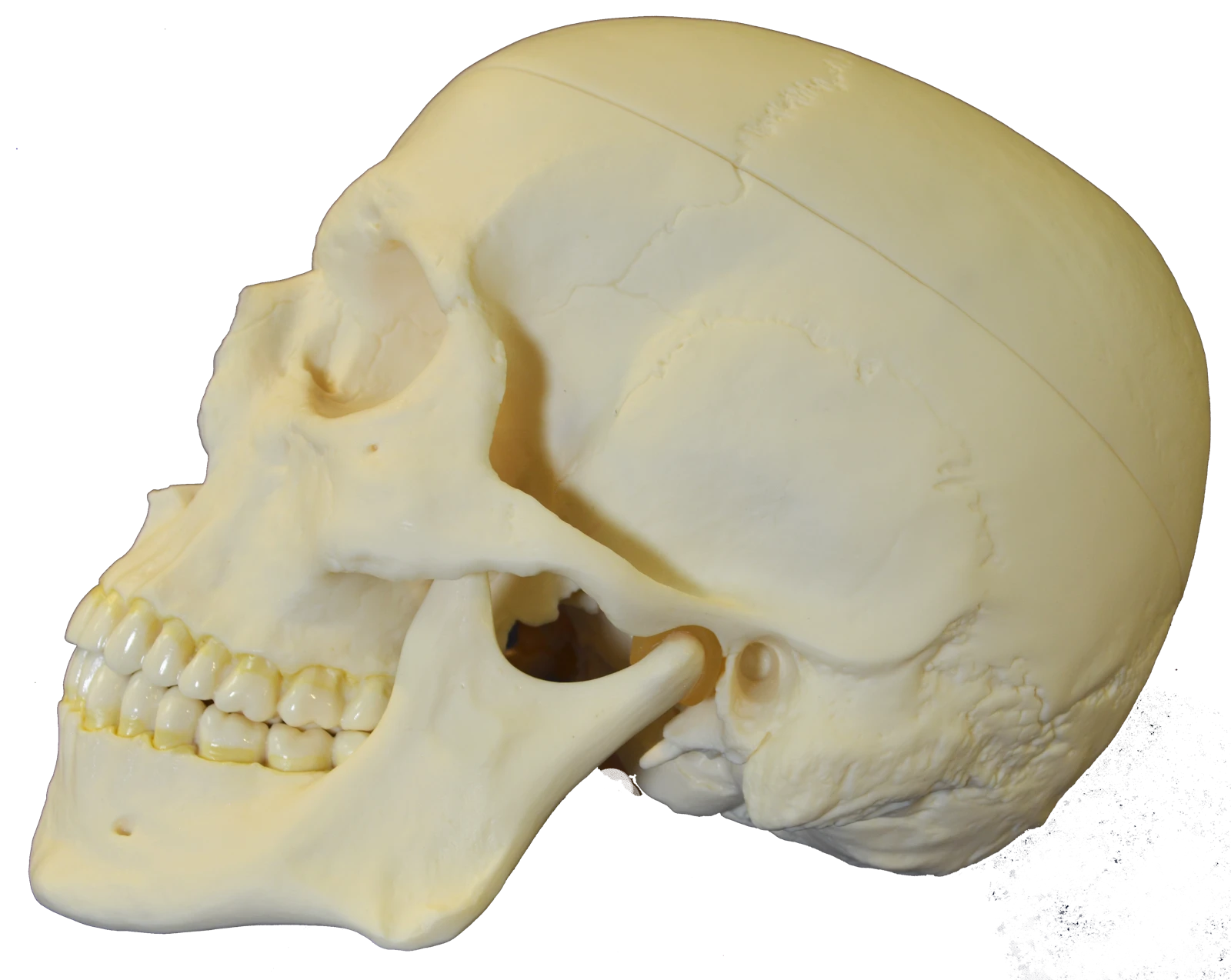
The mandible supports the lower teeth. Its condylar head articulates with the temporal bone at the temporomandibular joint.
This bony arch is formed by processes of the zygomatic and temporal bones.
This opening in the temporal bone leads into the external acoustic canal. It lies posterior to the mandibular condyle.
Which bone supports the lower teeth and includes the region marked A?
正解:D. Mandible
Aは下顎骨の骨体。下顎骨は下の歯を支え、下顎頭で側頭骨と顎関節を作る。上顎骨は上の歯を支える。頬骨は頬部、頭頂骨は頭蓋冠、側頭骨は頭蓋の側方に位置する。
A lies on the body of the mandible, which supports the lower teeth and articulates with the temporal bone through its condylar head. The maxilla supports the upper teeth. The zygomatic bone forms the cheek, the parietal bone contributes to the cranial vault, and the temporal bone lies at the side of the skull.
下顎骨を前方の骨体から後方の下顎枝へたどり、頬骨弓や外耳道との位置関係を確認する。
Trace the mandibular body back to the ramus, then compare its position with the zygomatic arch and external acoustic meatus.
図版:Bruce Forciea (PhysioAnatomica) · 出典 · CC BY-SA 4.0
原画像の構造・色・背景は変更せず、表示用に縮小してWebPへ変換。 目印・解説・問題はMIRUNOTEが追加。
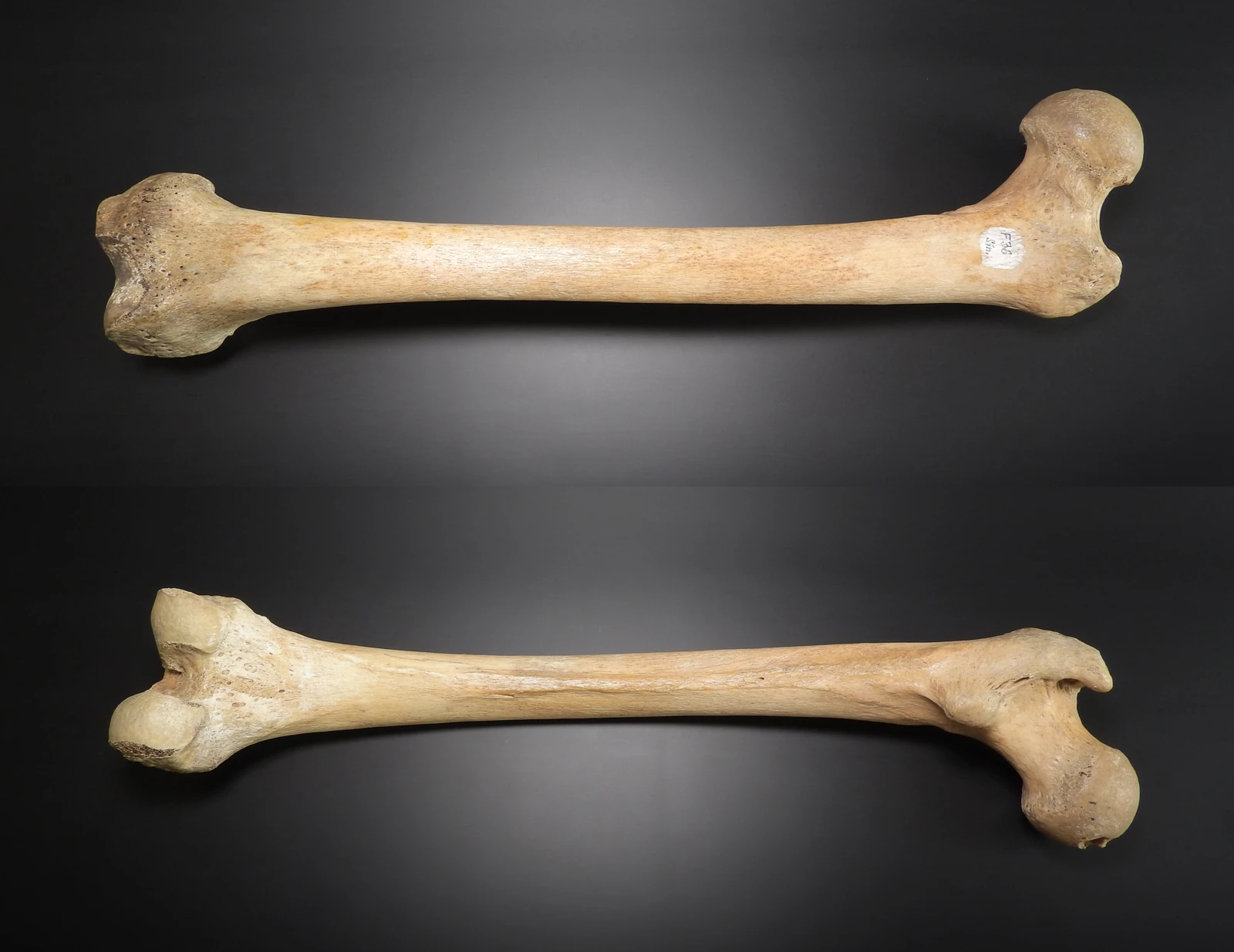
図で練習する ↑The spherical articular surface at the proximal end of the femur fits into the acetabulum to form the hip joint.
The narrower region connects the femoral head to the shaft region. It is distinct from the articular surface of the head.
This large lateral projection on the proximal femur provides attachment for muscles including gluteus medius and gluteus minimus.
The rounded articular surface marked A fits into which structure?
正解:B. Acetabulum
Aは大腿骨頭。寛骨臼と組み合わさって股関節を作る。肩甲骨の関節窩は上腕骨頭、尺骨の滑車切痕は上腕骨滑車と関節する。脛骨上面は膝関節の一部で、肘頭窩は上腕骨後面の窪み。
A marks the femoral head, which articulates with the acetabulum at the hip. The glenoid cavity receives the humeral head, and the trochlear notch receives the humeral trochlea. The tibial plateau participates in the knee joint, while the olecranon fossa is a depression on the posterior humerus.
大腿骨頭・頸・大転子を見分けた後、骨幹をたどって遠位端の顆を確認する。
Identify the head, neck and greater trochanter, then follow the shaft to the distal condyles.
原画像の構造・色・背景は変更せず、表示用に縮小してWebPへ変換。 目印・解説・問題はMIRUNOTEが追加。
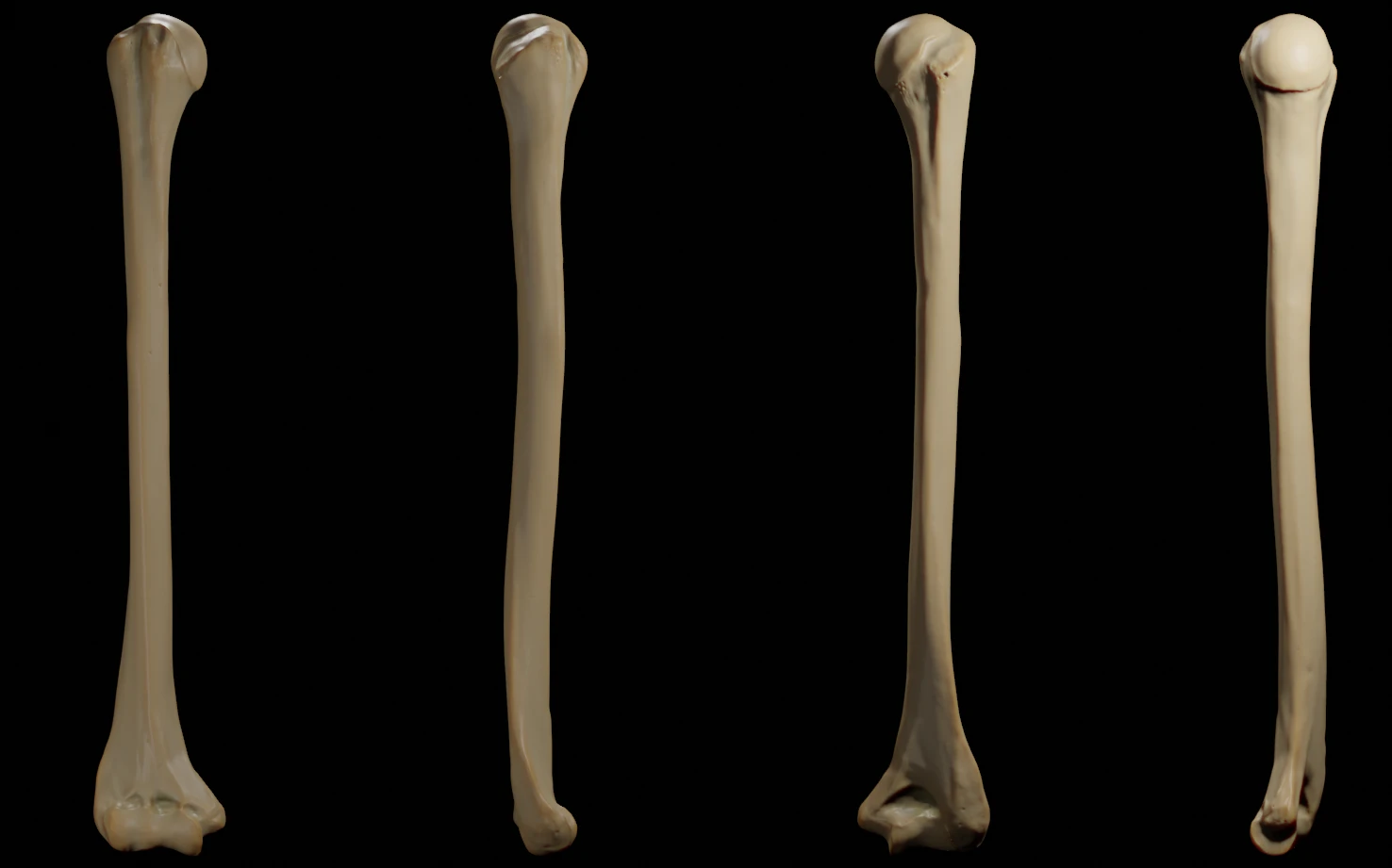
図で練習する ↑The rounded proximal articular surface meets the glenoid cavity of the scapula. It is visible in the rightmost view.
The shaft is the long central portion of the bone, between its proximal and distal ends.
This depression lies on the posterior distal humerus and receives the olecranon of the ulna during elbow extension.
Which bone articulates with the rounded articular surface marked A?
正解:C. Scapula
Aは上腕骨頭。肩甲骨の関節窩と肩関節を作る。橈骨・尺骨は上腕骨の遠位端と関節を作り、鎖骨・胸骨は上腕骨頭と直接関節を作らない。
A marks the head of the humerus. It articulates with the glenoid cavity of the scapula at the shoulder. The radius and ulna articulate with the distal humerus; the clavicle and sternum do not articulate directly with the humeral head.
骨頭がある近位端と、肘関節を作る遠位端を見分ける。
Distinguish the proximal humeral head from the distal surfaces that form the elbow joint.
図版:Doctor Jana · 出典 · CC BY-SA 4.0
解剖構造・組織像は変更せず、表示用にWebPへ変換。 目印・解説・問題はMIRUNOTEが追加。
図で練習する ↑英語の設問に答え、日本語・英語の解説で考え方を確認。
MIRUNOTE作成のオリジナル問題。
解き終わった問題の復習や、理由を確かめたいときに。各項目を開くと、設問・解答・解説・参考資料をまとめて読めます。
正解:B. Brachialis
上腕筋は上腕骨前面から尺骨に付着し、肘関節を屈曲させる。前腕の回内・回外にかかわらず肘屈曲に働く。
上腕三頭筋は肘伸展、棘上筋は肩外転、棘下筋は肩外旋に関与。広背筋は主に肩の伸展・内転・内旋に働く。
Brachialis runs from the anterior humerus to the ulna and is a primary elbow flexor. It contributes to flexion regardless of whether the forearm is pronated or supinated.
Triceps brachii extends the elbow. Supraspinatus contributes to shoulder abduction, infraspinatus to lateral rotation, and latissimus dorsi mainly to shoulder extension, adduction and medial rotation.
正解:D. Pulmonary trunk
右室から出た血液は肺動脈幹を通り、左右の肺動脈へ進む。「動脈」は心臓から出る方向で定義され、酸素量だけで分類するわけではない。
肺静脈は肺から左房へ戻る血管。大動脈は左室から体循環へ出る血管。上大静脈は上半身から右房へ戻る血管。
Blood leaves the right ventricle through the pulmonary trunk, which divides into the right and left pulmonary arteries. An artery carries blood away from the heart; its name does not depend on oxygen content.
Pulmonary veins return blood from the lungs to the left atrium. The aorta leaves the left ventricle for the systemic circulation. The superior vena cava returns blood from the upper body to the right atrium.
大学の試験が骨・筋・神経の同定を重視するのか、器官の位置関係や発生まで問うのかを確認します。講師が図の向きと構造間の関係を解説し、別の図や標本でも同じ部位を識別できるよう指導します。
授業では、自分で思い出す、図にする、問いに答える時間を設けます。講師と考え方を確かめ、説明しきれなかった部分を学び直してから、条件を変えた問題に取り組みます。
骨の目印、関節の動き、筋の起始・停止・作用を関連づけて学びます。筋の名前から動きを答えるだけでなく、動きから関わる筋や神経を考えます。
神経叢、末梢神経、主な動脈・静脈の走行と分布を学びます。どこを通り、何を支配・栄養するのかを、周囲の構造と一緒に整理します。
臓器、膜、腔、血管の位置関係を、表面の図と断面図で確かめます。隣り合う臓器や前後・上下の関係を、自分で図を描きながら説明します。
頭頸部の区画、脳神経の経路、感覚器の構造を学びます。解剖図・標本写真・断面画像を見比べ、向きが変わっても構造を捉える練習をします。
知識を思い出す練習と、その知識を使って考える練習を組み合わせます。解答だけでなく、そこに至る理由も講師と一緒に確かめます。
腕神経叢などを何も見ずに描き、神経のつながりと支配領域を説明します。講師と抜けを確かめて描き直し、別の部位でも同じ手順で整理します。
名称を伏せた図で、位置、形、隣接する構造を根拠に部位を答えます。正面図で答えられたら、断面や別方向の図へ進みます。
ある神経が障害された場合の運動・感覚の変化を考えます。答えを選んだ理由と、似た神経を候補から外す理由も説明します。
例:腕神経叢(Brachial plexus)。全体構成から末梢神経、支配領域へと追加質問を進めます。名称の列挙にとどまった回答には、どの位置関係・機能を補うべきか講師が示し、英語で回答を組み直します。
末梢神経損傷を題材に、症例で示された運動・感覚の情報から障害部位を検討する演習に取り組みます。講師が、似た神経や損傷部位を区別する所見を解説します。
上記は授業での学び方の例です。解説は日本語または英語で行い、口頭試験の練習では大学で使う英語の用語・表現も身につけます。
講義の復習、試験までの予定、留学生活の不安。現在の状況を伺い、必要な授業と相談の進め方をご案内します。